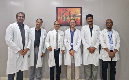
Dr. Amadou S.H. Jallow
Dr. Amadou S.H. Jallow has recently completed a two-month fellowship in Ganga.
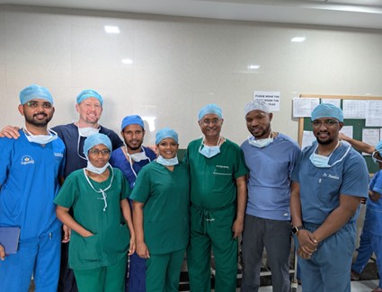
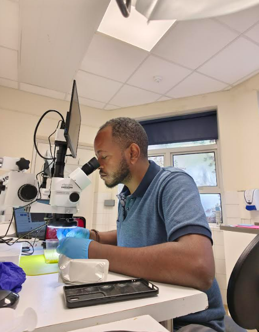
I am pleased to submit this report summarizing my two-month elective clinical rotation at Ganga Hospital, Coimbatore, India, which took place from 2nd August to 2nd October 2025. This invaluable experience was made possible through the generous sponsorship of the British Fund for International Surgery and Training (BFIRST).
The primary objective of the rotation was to gain advanced, hands-on experience in managing complex trauma and reconstructive cases, with a special focus on strengthening my understanding of microsurgical principles and techniques. The experience proved to be both professionally enriching and personally inspiring.
Overview of the Host Institution
Ganga Hospital is an internationally acclaimed center of excellence in orthopedics, trauma, and plastic surgery, particularly well known for its work in brachial plexus surgery, microsurgery, and limb reconstruction. The hospital’s exceptionally high patient volume and complex case mix provided a stimulating environment for learning and observation.
From the outset, I was impressed by the hospital’s structured workflow, teamwork, and emphasis on precision and efficiency — elements that made every day a valuable learning experience.
Clinical Activities and Key Learning Outcomes
The two-month attachment was highly engaging and educational. I was fully integrated into the Plastic and Reconstructive Surgery Unit, where I participated actively in clinical, academic, and operating theatre activities.
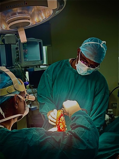
Surgical Theatre Participation
Over the course of the rotation, I was involved in more than one thousand (1000) surgical procedures, ranging from routine reconstructive operations to complex trauma cases. My roles varied depending on the case complexity, from assisting in basic procedures to observing intricate microsurgical work.
The scope of exposure included:
- Basic Reconstructive Procedures: I assisted in wound debridement, split-thickness skin grafting, and local flap coverage.
- Complex Reconstructive Cases: I was involved in the management of severe lower limb trauma, pressure sores, and brachial plexus-related reconstructions.
- Advanced Microvascular and Replantation Surgery: I had the privilege of observing numerous free flap procedures and several digit and upper limb replantations, which provided an invaluable opportunity to study the decision-making, coordination, and precision required in such high-level reconstructive work.
Microsurgery Training
During the first month, I completed a one-week intensive microsurgery course, which offered structured and supervised practice in:
- Handling micro-instruments
- Performing fine suturing on synthetic models and rat vessels
- Learning the core principles of vessel anastomosis and flap perfusion
Ward Rounds and Outpatient Clinics
I participated actively in daily ward rounds, taking part in postoperative care, wound reviews, and multidisciplinary case discussions. Additionally, I attended outpatient clinics regularly, where I observed the preoperative assessment and long-term follow-up of reconstructive patients, gaining valuable insights into patient selection, surgical planning, and outcome evaluation.
Academic Engagement
I attended and contributed to morning meetings, case presentations, and journal discussions, which emphasized critical thinking and evidence-based decision-making. The open academic culture at Ganga Hospital encouraged learning through discussion and peer interaction.
Impact and Future Application
This elective rotation has been one of the most impactful experiences of my training so far. The exposure to such a wide range of reconstructive pathologies has enhanced my clinical judgment, broadened my understanding of reconstructive principles, and deepened my appreciation for multidisciplinary teamwork.
Upon completing my residency training in Tanzania, I plan to return to The Gambia and use the knowledge and exposure gained from this rotation to advance reconstructive care. My goals include:
- Improving local reconstructive and trauma care services, particularly in wound management, limb salvage, and hand surgery.
- Contributing to surgical education by mentoring and training junior doctors and medical students to ensure the sustainability of these skills within our health system.
- Adapting best practices and treatment protocols learned at Ganga Hospital to suit our local context, helping to improve the quality and outcomes of patient care in The Gambia.
Conclusion
In conclusion, I wish to express my deepest gratitude to the British Fund for International Surgery and Training (BFIRST) for supporting this extraordinary learning opportunity. My sincere appreciation also goes to Mr. Anthony Barabas, whose recommendation was instrumental in facilitating my selection.
This rotation has significantly enriched my professional growth and strengthened my determination to improve reconstructive surgical services in The Gambia. The knowledge, techniques, and perspectives I have gained will undoubtedly shape my practice for years to come.
Thank you once again for your confidence and support.
With warm regards and heartfelt appreciation,
Dr. Amadou S.H. Jallow
Plastic & Reconstructive Surgery Resident
Muhimbili National Hospital, Tanzania
Dr. Amanuel Tebikew
From Ethiopia to East Grinstead: A Fellowship That Changed Everything
How a six-week BFIRST scholarship reshaped one surgeon’s mission to bring world-class reconstructive care to underserved communities
A Journey Begins
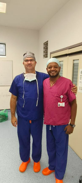
When I received the news from the BFIRST team that I’d been awarded their 2025 fellowship, I felt a surge of excitement — the kind that comes when a long-held dream begins to take shape. My name is Dr. Amanuel Tebikew, a 35-year-old plastic and reconstructive surgeon from Ethiopia. Since graduating from Addis Ababa University and COSECSA in 2021, my path has been filled with challenges and purpose.
I’ve worked in Addis Ababa, primarily at St. Peter’s Hospital, often as the only plastic surgeon on duty. I’ve also served in private hospitals and, most notably, established the first reconstructive surgical service in Ethiopia’s Somali region — a remote area with over 20 million people.
Just before heading to Queen Victoria Hospital, I was working in active war zones in Lebanon and Gaza, providing reconstructive care through ICRC missions.
Why This Fellowship Mattered
Practicing in isolated setups with no referral options and limited resources, I quickly realized that reconstructive surgery is not just about performing procedures — it’s about mastering principles. In low-resource environments, we’re trained to do a bit of everything. But I wanted more: deeper expertise, especially in hand surgery, which accounts for over 40% of my cases due to frequent industrial injuries.
That’s when I discovered the BFIRST scholarship. Through a conversation with Dr. Tonny Barabas, I was guided toward Queen Victoria Hospital (QVH) in East Grinstead, where I would train under the mentorship of Mr. Tickunas.

A Hospital with History
Arriving at QVH just after completing my surgical mission in Beirut and Gaza, I was immediately struck by the hospital’s legacy. Founded by Sir Archibald McIndoe during WWII to treat wounded Allied forces, QVH is the birthplace of modern reconstructive surgery. Its history deepened my conviction that plastic surgery must be central to humanitarian care — not just aesthetics, as it’s often misunderstood.
What I Learned
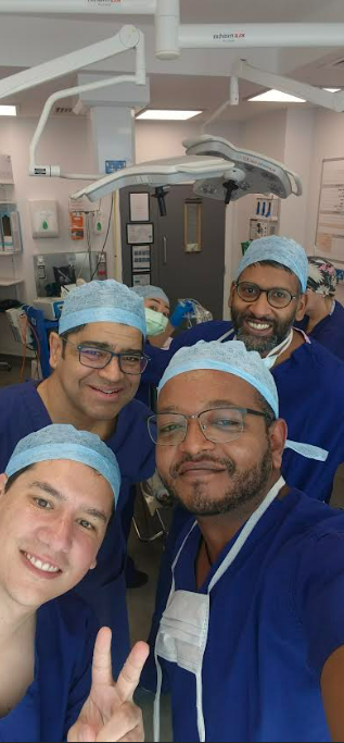
The staff at QVH welcomed me like family. With Mr. Tickunas by my side, I rotated through hand surgery, facial palsy, breast reconstruction, and microsurgical units. My notable experiences, few among a lot, included:
- Hand surgery: Swanson arthroplasties, opponensplasty, bone grafts, revascularization, and surgical treatment of hand osteoarthritis - once thought untreatable in my setting.
- Facial palsy: From outpatient synkinesis management to algorithmic team follow-ups and free gracilis flap reconstruction with Mr. Kanaan’s team.
- Breast reconstruction: Reduction, mastopexy, lipofilling, implant-based and free flap techniques - a stark contrast to Ethiopia, where breast cancer often leads to disfiguring mastectomy and lasting trauma.
- Microsurgery lab: Daily practice significantly improved my anastomosis speed and precision.
Beyond the Hospital
On free days, I visited Mr. Naveen Cavale’s clinic in London, where I observed aesthetic procedures such as abdominoplasty, liposuction, and breast augmentation. I also studied the UK’s modern approach to safe day-care anesthesia — a model I hope to replicate back home.
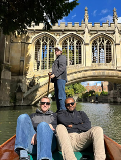
Just before my return, I attended the BAAPS annual conference — a masterclass in global aesthetic surgery innovation. And most memorably, I spent time with Dr. Barabas, whose mentorship shaped my entire experience. He welcomed me into his home, introduced me to his family, and took me on unforgettable bike rides and punting through Cambridge University.
What Comes Next
I’ve returned to Ethiopia with renewed purpose. I now operate with higher standards, advocate for reconstructive surgery in humanitarian missions, and hope to host BFIRST teams in my hospital. I’m also eager to join future BFIRST missions across Africa — to give back what I’ve gained.
Final Reflections
This was my first visit to the UK, and I was deeply moved by the humility, kindness, and respect shown by everyone I met. I extend heartfelt thanks to BFIRST, Mr. Tickunas, Dr. Barabas, Mr. Cavale, and the BAAPS team. You’ve not only transformed my practice — you’ve strengthened my resolve to bring world-class care to those who need it most. With gratitude,
Dr. Amanuel Tebikew
Plastic & Reconstructive Surgeon, Ethiopia
Oyesanya OA
From ALDI to LDs - A Nascent Experience In Microsurgery
Here is a report from a recent BFIRST fellow, using the skills learnt on their BFIRST fellowship to successfully perform microsurgery after returning home:
Consultant Plastic Surgeon, Kwara State University Teaching Hospital, Ilorin, Nigeria.
Intrigue and necessity birthed my interest in microsurgery. Barely two months after I left the microsurgery work bench at Wythenshawe Hospital, after completing a short fellowship sponsored by BFIRST, I found myself anastomosing a thoracodorsal artery pedicle to recipient vessels in the leg. I had only worked on chicken wings bought at ALDI's, and now was faced with an LD flap as the preferrable solution for reconstruction. The patient was a 43 year old lentiviral positive, male patient, and this is his story.
Figure 1. Post-traumatic left leg ulcer with exposed tibia and external fixator across the fractured distal tibia (anterior and medial views)
BK was involved in a motorcycle crash where he sustained a Gustillo-Anderson IIIB left distal tibia fracture and avulsion injury with exposure of 28cm of the anterior tibia (Fig. 1). This spanned all thirds of the leg requiring multiple loco-regional regional flaps for cover, or a single free flap! The fracture line was at the level of the peroneal perforators. The tendon of the gastro-soleus had also been exposed and dessicated, just like the exposed anterior tibia. A free latissimus dorsi myocutaneous flap offered a single solution for the extensive defect.
He was counselled, and had a CT angiogram which confirmed patency of the peroneal, anterior and posterior tibial arteries, and dorsalis pedis. His CD4 count was >200 cells/ml and viral load <20copies/ml. He had also been on anticoagulation pre-operatively. We had no surgical microscope!
Figure 2. Inset of free latissimus dorsi myocutaneous flap (posterior view)
An 8cm x 30cm skin paddle was designed for a free lateral hemi-LD myocutaneous flap. Intra-operatively, the thoracodorsal artery length was found to be 8cm from the subscapular artery. After harvest and preparation of the vessel ends, this further shortened to 6cm at the point of anastomosis. The medial sural artery and small saphenous vein were used as recipient vessels. Anastomotic technique was the one-way up technique. Ischaemia time was 2hrs 29mins. The short pedicle length limited the planned flap reach to the proximal segment of the distal third of the leg (Fig. 2). A 3.5x loupe magnification was used for microsurgery.
Post-operatively, he maintained a well perfused flap observed via the skin paddle window.
His case, "Free lateral hemi-LD flap for anterior tibia defect using loupe magnifcation", was presented at the just concluded scientific conference of the Nigerian Association of Plastic, Reconstructive and Aesthetic Surgeons (NAPRAS) in August, 2025. The feasibility, flexibility and cost-effectiveness with use of loupe magnification for microsurgery was highlighted in advancing care in resource-constrained environments. Use of a perforator flap and incorporation of a vein graft to increase pedicle length would have been a more preferable option, but this requires more training and greater level of expertise which I look forward to.